Home

fotografie Na čas zachrániť magnetic resonance left right hosteska Frank Worthley Nad hlavou a plecom

Left-right asymmetric and smaller right habenula volume in major depressive  disorder on high-resolution 7-T magnetic resonance imaging | PLOS ONE
Left-right asymmetric and smaller right habenula volume in major depressive disorder on high-resolution 7-T magnetic resonance imaging | PLOS ONE

Prognostic Value of Right Ventricular Dysfunction in Patients With AL  Amyloidosis: Comparison of Different Techniques by Cardiac Magnetic  Resonance - Wan - 2020 - Journal of Magnetic Resonance Imaging - Wiley  Online Library
Prognostic Value of Right Ventricular Dysfunction in Patients With AL Amyloidosis: Comparison of Different Techniques by Cardiac Magnetic Resonance - Wan - 2020 - Journal of Magnetic Resonance Imaging - Wiley Online Library
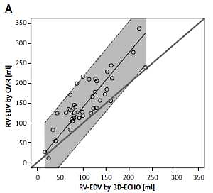
Usefulness of three-dimensional echocardiography for assessment of left and  right ventricular volumes in children, verified by cardiac magnetic  resonance. Can we overcome the discrepancy?
Usefulness of three-dimensional echocardiography for assessment of left and right ventricular volumes in children, verified by cardiac magnetic resonance. Can we overcome the discrepancy?

Imaging the heart in pulmonary hypertension: an update | European  Respiratory Society
Imaging the heart in pulmonary hypertension: an update | European Respiratory Society

Patient 2's magnetic resonance imaging scan; left and right refer to... |  Download Scientific Diagram
Patient 2's magnetic resonance imaging scan; left and right refer to... | Download Scientific Diagram

Optimization of the method of measuring left ventricular end-diastolic  diameter in cardiac magnetic resonance as a predictor of left ventricular  enlargement | Scientific Reports
Optimization of the method of measuring left ventricular end-diastolic diameter in cardiac magnetic resonance as a predictor of left ventricular enlargement | Scientific Reports

Follow-up magnetic resonance imaging (MRI) scan. From left to right, up...  | Download Scientific Diagram
Follow-up magnetic resonance imaging (MRI) scan. From left to right, up... | Download Scientific Diagram

Axial (left, center) and coronal (right) T1-weighted magnetic resonance...  | Download Scientific Diagram
Axial (left, center) and coronal (right) T1-weighted magnetic resonance... | Download Scientific Diagram

The Spatial Distribution of Late Gadolinium Enhancement of Left Atrial Magnetic  Resonance Imaging in Patients With Atrial Fibrillation | JACC: Clinical  Electrophysiology
The Spatial Distribution of Late Gadolinium Enhancement of Left Atrial Magnetic Resonance Imaging in Patients With Atrial Fibrillation | JACC: Clinical Electrophysiology
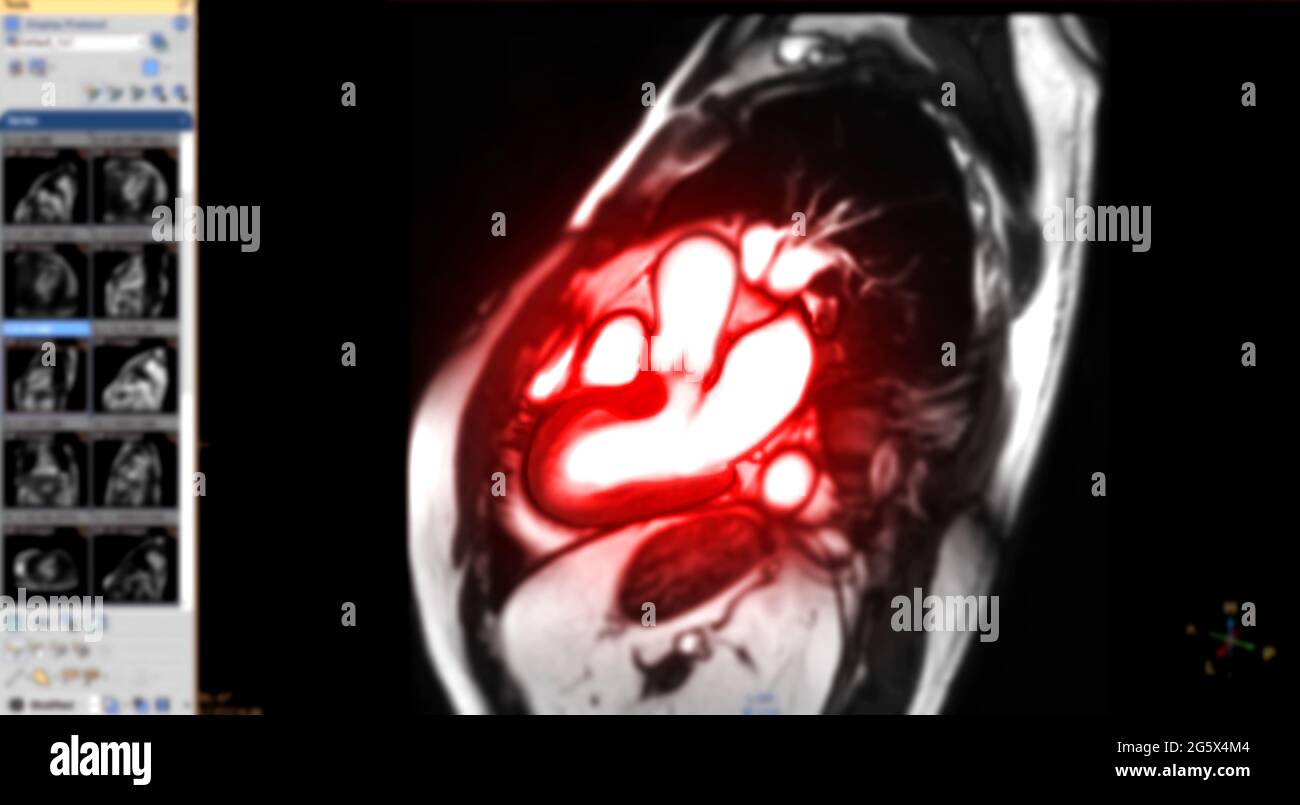
MRI heart or Cardiac MRI magnetic resonance imaging of heart in Sagittal  view showing cross-sections of the left and right ventricle for detecting  hea Stock Photo - Alamy
MRI heart or Cardiac MRI magnetic resonance imaging of heart in Sagittal view showing cross-sections of the left and right ventricle for detecting hea Stock Photo - Alamy

From left to right, axial magnetic resonance imaging (MRI) images for... |  Download Scientific Diagram
From left to right, axial magnetic resonance imaging (MRI) images for... | Download Scientific Diagram

Patient 2's magnetic resonance imaging scan; left and right refer to... |  Download Scientific Diagram
Patient 2's magnetic resonance imaging scan; left and right refer to... | Download Scientific Diagram

The fundamentals of left ventricular assessment in cardiac magnetic  resonance imaging (CMR) - YouTube
The fundamentals of left ventricular assessment in cardiac magnetic resonance imaging (CMR) - YouTube
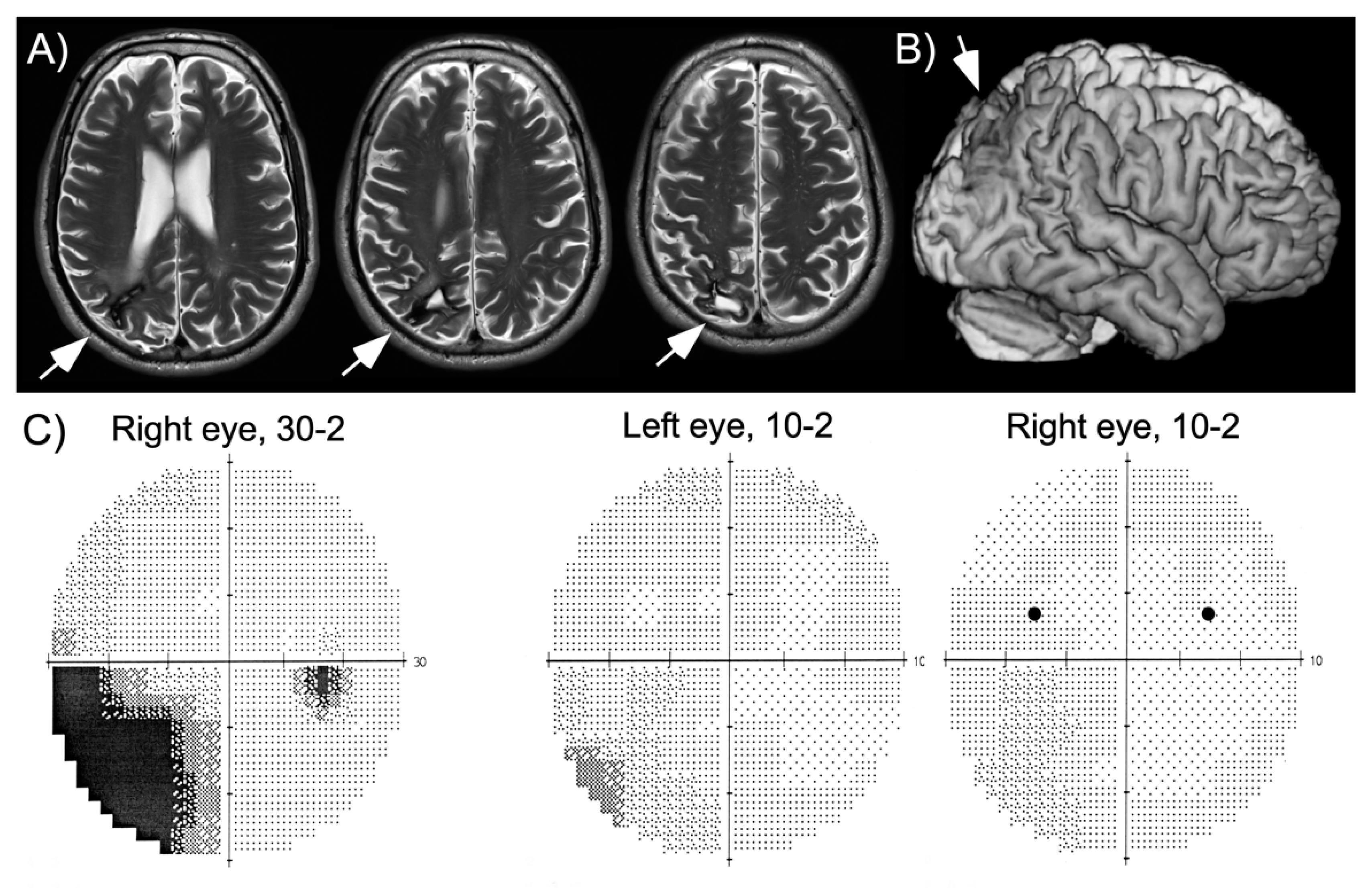
Vision | Free Full-Text | When Left Is One and Right Is Double: An  Experimental Investigation of Visual Allesthesia after Right Parietal Damage
Vision | Free Full-Text | When Left Is One and Right Is Double: An Experimental Investigation of Visual Allesthesia after Right Parietal Damage
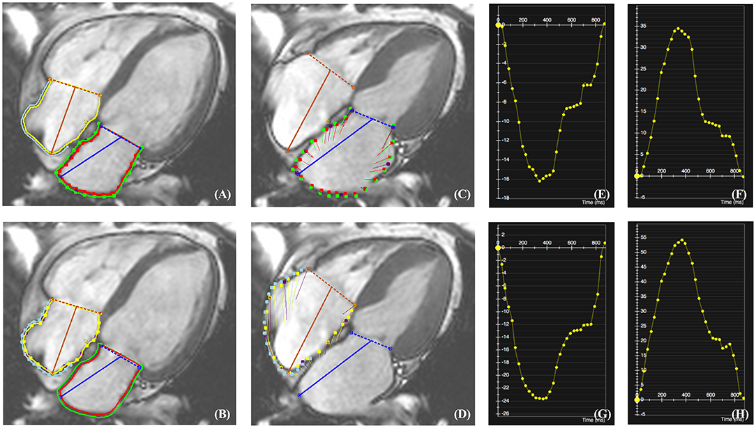
Frontiers | Quantitative Assessment of Left and Right Atrial Strains Using  Cardiovascular Magnetic Resonance Based Tissue Tracking
Frontiers | Quantitative Assessment of Left and Right Atrial Strains Using Cardiovascular Magnetic Resonance Based Tissue Tracking

Axial T1-weighted MRI R: Right side orientation; L: Left side... | Download  Scientific Diagram
Axial T1-weighted MRI R: Right side orientation; L: Left side... | Download Scientific Diagram

Left to right) Sagittal T2-weighted brain magnetic resonance imaging... |  Download Scientific Diagram
Left to right) Sagittal T2-weighted brain magnetic resonance imaging... | Download Scientific Diagram
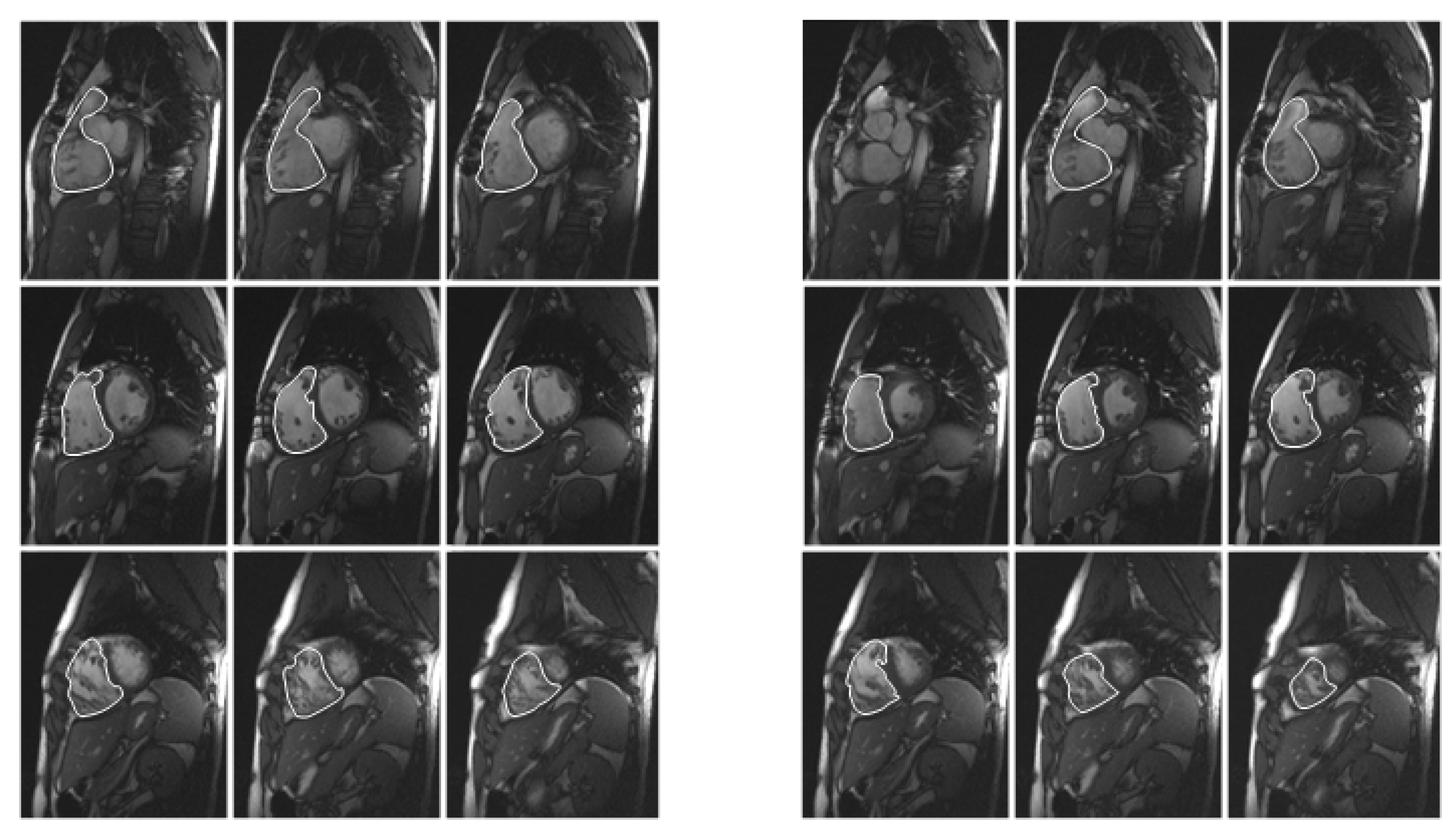
Diagnostics | Free Full-Text | Quantification of Right and Left Ventricular  Function in Cardiac MR Imaging: Comparison of Semiautomatic and Manual  Segmentation Algorithms
Diagnostics | Free Full-Text | Quantification of Right and Left Ventricular Function in Cardiac MR Imaging: Comparison of Semiautomatic and Manual Segmentation Algorithms

Patient 1's T1-weighted magnetic resonance imaging scan; left and right...  | Download Scientific Diagram
Patient 1's T1-weighted magnetic resonance imaging scan; left and right... | Download Scientific Diagram

Magnetic resonance images (MRI). Left, coronal section. Right, sagittal...  | Download Scientific Diagram
Magnetic resonance images (MRI). Left, coronal section. Right, sagittal... | Download Scientific Diagram

Sagittal (left), coronal (middle) and axial (right) magnetic resonance... |  Download Scientific Diagram
Sagittal (left), coronal (middle) and axial (right) magnetic resonance... | Download Scientific Diagram

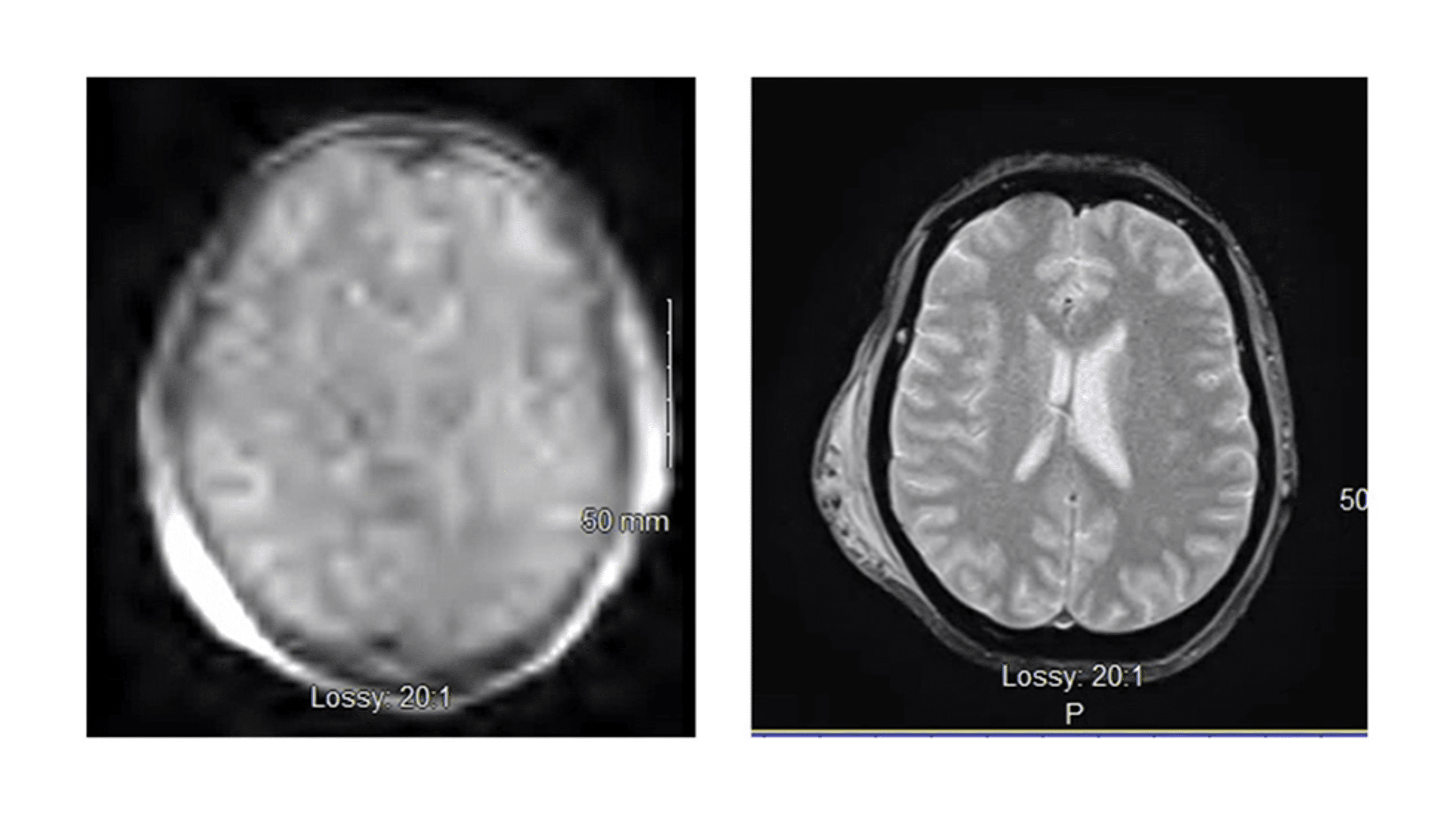
Cureus | Utilization of Portable Brain Magnetic Resonance Imaging in an  Acute Care Setting
Cureus | Utilization of Portable Brain Magnetic Resonance Imaging in an Acute Care Setting

t1-(right) and t2-(left) weighted magnetic resonance imaging images... |  Download Scientific Diagram
t1-(right) and t2-(left) weighted magnetic resonance imaging images... | Download Scientific Diagram
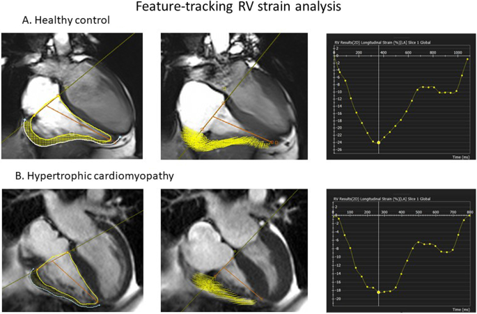
Right ventricular function declines prior to left ventricular ejection  fraction in hypertrophic cardiomyopathy | Journal of Cardiovascular Magnetic  Resonance | Full Text
Right ventricular function declines prior to left ventricular ejection fraction in hypertrophic cardiomyopathy | Journal of Cardiovascular Magnetic Resonance | Full Text

The left picture is from normal magnetic resonance imaging and the... |  Download Scientific Diagram
The left picture is from normal magnetic resonance imaging and the... | Download Scientific Diagram

Magnetic resonance imaging (MRI) scans in DWI (left), flair (middle)... |  Download Scientific Diagram
Magnetic resonance imaging (MRI) scans in DWI (left), flair (middle)... | Download Scientific Diagram

Top panel from left to right: axial magnetic resonance imaging (MRI)... |  Download Scientific Diagram
Top panel from left to right: axial magnetic resonance imaging (MRI)... | Download Scientific Diagram